Guidelines On How To Spot Heel Spur

Inferior Calcaneal Spur

Overview

Heel spurs are a common foot problem resulting from excess bone growth on the heel bone. The bone growth is usually located on the underside of the heel bone, extending forward to the toes. One explanation for this excess production of bone is a painful tearing of the plantar fascia connected between the toes and heel. This can result in either a heel spur or an inflammation of the plantar fascia, medically termed plantar fascitis. Because this condition is often correlated to a decrease in the arch of the foot, it is more prevalent after the age of six to eight years, when the arch is fully developed.

Causes

Each time we take a step forward, all of our body weight first rests on the heel of one foot. As our weight moves forward, the entire foot begins to bear the body’s weight, and the foot flattens and this places a great deal of pressure and strain on the plantar fascia. There is very little ?give? to the plantar fascia, so as it stretches only slightly, it pulls on its attachment to the heel. If the foot is properly aligned this pull causes no problems. However, if the foot is ?pronated?(the foot rolls outward at the ankle, causing a break down of the inner side of the shoe), the arch falls excessively, and this causes an abnormal stretching of the relatively inflexible plantar fascia, which in turn pulls abnormally hard on the heel. The same pathology occurs with ?supination? (the rolling inward of the foot, causing a break down of the outer side of the shoe). Supinated feet are relatively inflexible; usually have a high arch, and a short or tight plantar fascia. Thus as weight is transferred from the heel to the remainder of the foot, the tight plantar fascia hardly stretches at all, and pulls with great force on its attachment to the heel. In both cases, the abnormal stress placed on the attachment of the plantar fascia to the heel usually causes pain, inflammation, and possibly swelling. If this process continues, the plantar fascia partially tears away from the heel. The body will fill in this torn area with calcium; eventually it becomes bone, and a heel spur results.
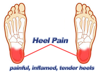
Inferior Calcaneal Spur

Symptoms

Most people think that a bone “spur” is sharp and produces pain by pressing on tissue, when in fact, these bony growths are usually smooth and flat. Although they rarely cause pain on their own, bone spurs in the feet can lead to callus formation as tissue builds up to provide added cushion over the area of stress. Over time, wear and tear on joints may cause these spurs to compress neighboring ligaments, tendons or nerves, thus injuring tissue and causing swelling, pain and tearing.

Diagnosis

Your doctor will discuss your medical history and will examine your foot and heel for any deformities and inflammation (swelling, redness, heat, pain). He/she will analyze your flexibility, stability, and gait (the way you walk). Occasionally an x-ray or blood tests (to rule out diseases or infections) may be requested.

Non Surgical Treatment

Bone spurs rarely require treatment unless they are causing frequent pain or damaging other tissues. Because heel spurs and plantar fasciitis are so closely related, they are usually treated the same way. Symptomatic treatment involves rest, especially from the activity that is contributing to the condition and making symptoms worse (although this may not be easy to discover, as problems can manifest several hours or days after the harmful activity has occurred). If you identify the offending activity, ice is recommended immediately following it. Stretching of the calf muscles after a short warm up is also a good idea and can be helpful. Stretching exercises that gently lengthen the calm muscle will relax the tissue surrounding the heel and should be done several times a day, especially in the morning and after prolonged sitting.

Surgical Treatment

Heel spur surgery should only be considered after less invasive treatment methods have been explored and ruled insufficient. The traditional surgical approach to treating heel spurs requires a scalpel cut to the bottom of the food which allows the surgeon to access the bone spur. Endoscopic plantar fasciotomies (EPF) involve one or two small incisions in the foot which allow the surgeon to access and operate on the bone spur endoscopically. Taking a surgical approach to heel spur treatment is a topic to explore with a foot and ankle specialist.

Therapy For Bursitis Of The Foot

Overview

Infracalcaneal bursitis can significantly affect your quality of life and your ability to perform your activities of daily living, due to pain and impaired gait. Inflammation of the bursal sac under your heel bone occurs because the bursa is abnormally stressed or strained in some way or bears excessive pressure for prolonged periods. Constant pressure and friction from footwear is a common cause of this health problem, and any treatment plan addressing infracalcaneal bursitis should include recommendations for footwear to avoid or use. Infracalcaneal bursitis may be diagnosed in several ways, including palpation, or light pressure applied to your affected area. If your heel pain has existed for an extended period, X-ray imaging studies may reveal localized calcification in your infracalcaneal bursa, though this is not always the case. MRI images are sometimes used as a diagnostic tool for this health problem, though MRI studies are considered unnecessary for diagnosis in many cases.

Causes

Bursitis occurs when the synovial lining becomes thickened and produces excessive fluid, leading to localized swelling and pain. It most commonly affects the subacromial, olecranon, trochanteric, prepatellar, and infrapatellar bursae. Symptoms of bursitis may include localized tenderness, pain, edema, erythema, or reduced movement. Pain is aggravated by movement of the specific joint, tendon, or both.

Symptoms

Limping. Decreased movement. Your ankles may feel stiff or unable to move as well as they usually do. Pain or tenderness in the back of the ankle. It may be worse at the beginning of exercise, or when running uphill. You may also have pain when wearing shoes. Redness and warmth. If the bursa is infected, the skin over the heel may be red and warm. You may also have a fever. Swelling on the back of the heel.

Diagnosis

Medical examination is not necessarily required in light cases where the tenderness is minimal. In all cases where smooth improvement is not experienced, medical attention should be sought as soon as possible to exclude a (partial) rupture of the Achilles tendon or rupture of the soleus muscle. This situation is best determined by use of ultrasound scanning, as a number of injuries requiring treatment can easily be overlooked during a clinical examination (Ultrasonic image). Ultrasound scanning enables an evaluation of the extent of the change in the tendon, inflammation of the tendon (tendinitis), development of cicatricial tissue (tendinosis), calcification, inflammation of the tissue surrounding the tendon (peritendinitis), inflammation of the bursa (bursitis), as well as (partial) rupture.

Non Surgical Treatment

Over-the-counter or custom heel wedges may help to decrease the stress placed on the attachment of the achilles tendon and the associated bursa. If these interventions are ineffective, then some health care providers may inject a small amount of steroids into the bursa. If the condition is associated with Achilles tendonitis, then casting the ankle to prevent motion for several weeks can be effective. Very rarely, surgery may be necessary to remove the inflamed bursa.

Surgical Treatment

Surgery is rarely need to treat most of these conditions. A patient with a soft tissue rheumatic syndrome may need surgery, however, if problems persist and other treatment methods do not help symptoms.

Prevention

Maintain proper form when exercising, as well as good flexibility and strength around the ankle to help prevent this condition. Proper stretching of the Achilles tendon helps prevent injury.

What Are The Causes Of Hammertoes

Hammer ToeOverview

hammertoes and mallet toe are two foot deformities that occur most often in women who wear high heels or shoes with a narrow toe box. These types of footwear may force your toes against the front of the shoe, causing an unnatural bending. A hammertoe has an abnormal bend in the middle joint of a toe. Mallet toe affects the joint nearest the toenail. Hammertoe and mallet toe are most likely to occur in the toe next to your big toe. Relieving the pain and pressure of hammertoe and mallet toe may involve changing your footwear and wearing shoe inserts. If you have a more severe case of hammertoe or mallet toe, you may need surgery to experience relief.

Causes

Hammer toes result from a muscle imbalance which causes the ligaments and tendons to become unnaturally tight. This results in the joint curling downward. Arthritis can also lead to many different forefoot deformities, including hammer toes.

HammertoeSymptoms

The symptoms of a hammer toe are usually first noticed when a corn develops on the top of the toe and becomes painful, usually when wearing tight shoes. There may be a bursa under hammertoe the corn or instead of a corn, depending on the pressure. Most of the symptoms are due to pressure from footwear on the toe. There may be a callus under the metatarsal head at the base of the toe. Initially a hammer toe is usually flexible, but when longstanding it becomes more rigid.

Diagnosis

The exam may reveal a toe in which the near bone of the toe (proximal phalanx) is angled upward and the middle bone of the toe points in the opposite direction (plantar flexed). Toes may appear crooked or rotated. The involved joint may be painful when moved, or stiff. There may be areas of thickened skin (corns or calluses) on top of or between the toes, a callus may also be observed at the tip of the affected toe beneath the toenail. An attempt to passively correct the deformity will help elucidate the best treatment option as the examiner determines whether the toe is still flexible or not. It is advisable to assess palpable pulses, since their presence is associated with a good prognosis for healing after surgery. X-rays will demonstrate the contractures of the involved joints, as well as possible arthritic changes and bone enlargements (exostoses, spurs). X-rays of the involved foot are usually performed in a weight-bearing position.

Non Surgical Treatment

If the problem is caught in the early stages you can avoid hammer toe surgery. One of the easiest methods of treatment is to manipulate the toe out of a bent position then splint and buddy wrap it alongside it?s larger neighbour. This method of hammer toe taping will help the problem to fix itself. Make sure the toe isn?t resuming its bent shape during the recovery. To alleviate some of the painful symptoms of hammer toe avoid wearing high heels or shoes that cramp or stifle your feet. Choosing a pair of minimalist shoes can be an excellent choice for both foot and postural health. Wearing shoes that give the toes plenty of space and are comfortable lined is also a smart choice. Hammer toe recovery starts be treating the toe respectfully. Soft insoles or protection for the corn can also provide additional assistance.

Surgical Treatment

Until recently, wires were used for surgical correction. In this technique, one or more wires are inserted into the bone through both the affected joint and a normally healthy toe joint, and the end of the toe. These wires stay in place for four to six weeks, protruding from the end of the toes. Due to the protruding wire, simple things such working, driving, bathing and even sleeping are difficult while these wires are in place. During this recovery period, patients often experience discomfort during sleep and are subject possible infection.

Hammer ToePrevention

Although the feet naturally change over time, and abnormalities like hammertoes may be hereditary for some patients, steps may be taken to prevent their development in the first place. Just as better fitting shoes are a treatment, they are also a preventative measure for hammertoes. In addition, your podiatrist may suggest orthotics to improve the biomechanics of your feet in an effort to prevent the development of hammertoes or other abnormalities. Calf stretching and other exercises may also be used to reverse or treat muscle imbalances that could eventually lead to hammertoe development.

What Are The Most Obvious Chief Reasons For Overpronation Of The Foot

Overview

Pronation is the normal movement the foot makes to absorb the impact from walking or running. It occurs once the heel strikes the ground and the foot disperses the impact, stretching and flattening the arch as the foot rolls inward. Supination is the opposite motion of pronation. The foot supinates, or rolls on its outer edge, to help with stability as we walk or run. A reasonable amount of pronation is necessary for the foot to function properly. However, when the foot arch remains flat and the foot rolls inward too much one may have excessive pronation or overpronation. This medical condition can result from continually straining the feet and wearing footwear that lacks sufficient foot arch support.Over-Pronation

Causes

A common cause of pronation is heredity – we can inherit this biomechanical defect. The second most common cause is due to the way our feet were positioned in the uterus while we were developing; this is called a congenital defect. In either instance, the following occurs in our feet during our development.

Symptoms

Eventually, over-pronation can lead to a full list of maladies including flat feet, plantar fasciitis, plantar fibroma, neuromas, heel spurs, shin splints, ankle sprains, bunions, hammertoes, calluses, and pain in the arches, knee, hip and lower back. But it doesn?t have to go that far, because there are steps we can take to correct the over-pronation. In the vast majority of cases, we?ll prescribe custom foot orthotics, which will realign your ankles, redistribute the weight, support the arch and reduce the twisting. Many orthotics will fit snugly into your normal shoes. Although we?ll also take a look at the type of shoes you wear to see if they are contributing to the problem.

Diagnosis

Bunions, calluses and crooked toes may indicate alignment problems. So, it is important to ascertain the condition of a client’s toes. Check the big toe to determine if the first joint of the toe is swollen, has a callus or bunion, and/or looks as though it abducts (i.e., hallux valgus) rather than pointing straight ahead. Also, look to see if the lesser toes seem to “curl up” (i.e., the person has hammer or claw toes). This may be indicative of damage to, or inflexibility of the plantar fascia caused by excessive flattening of the foot.Overpronation

Non Surgical Treatment

If a young child is diagnosed with overpronation braces and custom orthotics can be, conjunction with strengthening and stretching exercises, to realign the bones of the foot. These treatments may have to continue until the child has stopped growing, and orthotics may need to be worn for life in order to prevent the foot reverting to an overpronated state. Wearing shoes that properly support the foot, particularly the arch, is one of the most effective treatments for overpronation. Custom-made orthotic inserts can also be very beneficial. They too support the arch and distribute body weight correctly throughout the foot. Motion-control shoes that prohibit pronation can be worn, so may be useful for those with severe overpronation. One good treatment is to walk barefoot as often as possible. Not relying on shoes to support the arch will encourage proper muscle use. Practicing yoga can help to correct poor posture and teach you how to stand with your weight balanced evenly across the whole foot.

Prevention

Exercises to strengthen and stretch supporting muscles will help to keep the bones in proper alignment. Duck stance: Stand with your heels together and feet turned out. Tighten the buttock muscles, slightly tilt your pelvis forwards and try to rotate your legs outwards. You should feel your arches rising while you do this exercise. Calf stretch: Stand facing a wall and place hands on it for support. Lean forwards until stretch is felt in the calves. Hold for 30 seconds. Bend at knees and hold for a further 30 seconds. Repeat 5 times. Golf ball: While drawing your toes upwards towards your shins, roll a golf ball under the foot between 30 and 60 seconds. If you find a painful point, keep rolling the ball on that spot for 10 seconds. Big toe push:

Stand with your ankles in a neutral position (without rolling the foot inwards). Push down with your big toe but do not let the ankle roll inwards or the arch collapse. Hold for 5 seconds. Repeat 10 times. Build up to longer times and fewer repetitions. Ankle strengthener: Place a ball between your foot and a wall. Sitting down and keeping your toes pointed upwards, press the outside of the foot against the ball, as though pushing it into the wall. Hold for 5 seconds and repeat 10 times. Arch strengthener: Stand on one foot on the floor. The movements needed to remain balanced will strengthen the arch. When you are able to balance for 30 seconds, start doing this exercise using a wobble board.

Severs Disease Physiotherapy

Overview

Sever?s disease is caused by the growth plate in the heel becoming inflamed, and it is the most common cause of heel pain in adolescents. This condition is especially prevalent in children who play sports. Treatment includes ice, rest, and pain relievers to manage pain and discomfort. Any underlying foot conditions may also need to be assessed and managed. Sever?s disease does not cause any permanent damage, and will resolve when the growth of the heel is complete. Sever?s disease (also called calcaneal apophysitis) is a condition that occurs in the growth plate of the heel bone (the calcaneus) in children and adolescents. When the muscles and tendons in the leg and heel exert too much pressure on this growth plate, swelling and pain can result.

Causes

The spontaneous development of pain in children generally indicates some form of injury to the growth plate of a growing bone. This can occur without a specific memorable event. When pain occurs in the heel of a child the most likely cause is due to injury of the growth plate in the heel bone. This is called Sever’s disease. A condition that may mimic Seiver’s disease is Achilles tendonitis. Achilles tendonitis is inflammation of the tendon attached to the back of the heel. A tight Achilles tendon may contribute to Sever’s disease by pulling excessively on the growth plate of the heel bone. It is frequently seen in the active soccer, football or baseball player. Sport shoes with cleats seem to aggravate the condition. It is believed that the condition is due to an underlying mechanical problem with the way the foot functions.

Symptoms

The main symptom of sever’s disease is pain and tenderness at the back of the heel which is made worse with physical activity. Tenderness will be felt especially if you press in or give the back of the heel a squeeze from the sides. There may be a lump over the painful area. Another sign is tight calf muscles resulting with reduced range of motion at the ankle. Pain may go away after a period of rest from sporting activities only to return when the young person goes back to training.

Diagnosis

Physical examination varies depending on the severity and length of involvement. Bilateral involvement is present in approximately 60% of cases. Most patients experience pain with deep palpation at the Achilles insertion and pain when performing active toe raises. Forced dorsiflexion of the ankle also proves uncomfortable and is relieved with passive equinus positioning. Swelling may be present but usually is mild. In long-standing cases, the child may have calcaneal enlargement.

Non Surgical Treatment

Sever?s disease is a self-limiting problem, because as your child grows the growth plate will eventually fuse with the main body of the heel bone. This happens at about 14 -15 years of age. Once foot growth is complete and the growth plate has fused, the symptoms will resolve. In the meantime, treatment by your Podiatrist will help your child return to normal sporting activities without heel pain slowing him/her down.

Surgical Treatment

The surgeon may select one or more of the following options to treat calcaneal apophysitis. Reduce activity. The child needs to reduce or stop any activity that causes pain. Support the heel. Temporary shoe inserts or custom orthotic devices may provide support for the heel. Medications. Nonsteroidal anti-inflammatory drugs (NSAIDs), such as ibuprofen, help reduce the pain and inflammation. Physical therapy. Stretching or physical therapy modalities are sometimes used to promote healing of the inflamed issue. Immobilization. In some severe cases of pediatric heel pain, a cast may be used to promote healing while keeping the foot and ankle totally immobile. Often heel pain in children returns after it has been treated because the heel bone is still growing. Recurrence of heel pain may be a sign of calcaneal apophysitis, or it may indicate a different problem. If your child has a repeat bout of heel pain, be sure to make an appointment with your foot and ankle surgeon.

Heel Discomfort Everything It Is Best To Know Heel Painfulness

Overview

Heel Pain

Heel Pain is one of the most common conditions treated by podiatrists. It is often a message from the body that something is in need of medical attention. Pain that occurs right after an injury or early in an illness may play a protective role, often warning us about the damage we have suffered. The greatest incidence of heel pain is seen in middle-aged men and women. It is also seen in those who take part in regular sporting activities and those significantly overweight and on their feet a lot. Heel pain can also occur in children, usually between 8 and 13, as they become increasingly active in sporting activities.

Causes

he most common cause of heel pain is over pronation, this is when your foot rotates in too much as you walk. You really need to treat the underlying cause of the heel pain as soon as possible to prevent any further damage to the plantar fascia. Excessive load on the foot from obesity is a major cause of plantar fasciitis. Which is why this condition is common in middle aged and over weight adults. A sudden increase in weight, such as pregnancy can also lead to plantar fascitis. A sudden increase in walking or a sporting activity can also be a contributing factor. A classic example of when this condition can develop is when a post man has returned to work after a period away from the job. Tight plantar fascia (this is often caused by tight calf muscles). Excessive flattening of the arch on weight bearing i.e. flat feet. People with flat feet are more at risk of developing this condition. Biomechanical problems (walking abnormalities) is a major cause of plantar fasciitis. Different types of arthritis can also lead to this condition, such as osteoarthritis and rheumatoid arthritis.

Symptoms

Plantar fasciitis is a condition of irritation to the plantar fascia, the thick ligament on the bottom of your foot. It classically causes pain and stiffness on the bottom of your heel and feels worse in the morning with the first steps out of bed and also in the beginning of an activity after a period of rest. For instance, after driving a car, people feel pain when they first get out, or runners will feel discomfort for the first few minutes of their run. This occurs because the plantar fascia is not well supplied by blood, which makes this condition slow in healing, and a certain amount of activity is needed to get the area to warm up. Plantar fasciitis can occur for various reasons: use of improper, non-supportive shoes; over-training in sports; lack of flexibility; weight gain; prolonged standing; and, interestingly, prolonged bed rest.

Diagnosis

Your doctor will perform a physical exam and ask questions about your medical history and symptoms, such as have you had this type of heel pain before? When did your pain begin? Do you have pain upon your first steps in the morning or after your first steps after rest? Is the pain dull and aching or sharp and stabbing? Is it worse after exercise? Is it worse when standing? Did you fall or twist your ankle recently? Are you a runner? If so, how far and how often do you run? Do you walk or stand for long periods of time? What kind of shoes do you wear? Do you have any other symptoms? Your doctor may order a foot x-ray. You may need to see a physical therapist to learn exercises to stretch and strengthen your foot. Your doctor may recommend a night splint to help stretch your foot. Surgery may be recommended in some cases.

Non Surgical Treatment

Morning Wall Stretch. Stand barefoot in front of wall, as shown. Press into wall with both hands and lean forward, feeling stretch along back of left leg and heel. Hold for 30 seconds; switch sides and repeat. Freeze and Roll. Freeze a small water bottle. Cover it with a towel and place arch of your foot on top of it. Slowly roll bottle beneath arch of foot for about 5 minutes at a time. Switch sides and repeat. Rub It Out. Use both thumbs to apply deep pressure along arch of the feet, heel, and calf muscles, moving slowly and evenly. Continue for 1 minute. Switch sides and repeat. If you foot pain isn’t improving or worsens after 2 weeks, a podiatrist or othopedist can prescribe additional therapies to alleviate discomfort and prevent recurrence.

Surgical Treatment

Only a relatively few cases of heel pain require surgery. If required, surgery is usually for the removal of a spur, but also may involve release of the plantar fascia, removal of a bursa, or a removal of a neuroma or other soft-tissue growth.

Prevention

Pain Under The Heel

You should always wear footwear that is appropriate for your environment and day-to-day activities. Wearing high heels when you go out in the evening is unlikely to be harmful. However, wearing them all week at work may damage your feet, particularly if your job involves a lot of walking or standing. Ideally, you should wear shoes with laces and a low to moderate heel that supports and cushions your arches and heels. Avoid wearing shoes with no heels. Do not walk barefoot on hard ground, particularly while on holiday. Many cases of heel pain occur when a person protects their feet for 50 weeks of the year and then suddenly walks barefoot while on holiday. Their feet are not accustomed to the extra pressure, which causes heel pain. If you do a physical activity, such as running or another form of exercise that places additional strain on your feet, you should replace your sports shoes regularly. Most experts recommend that sports shoes should be replaced after you have done about 500 miles in them. It is also a good idea to always stretch after exercising, and to make strength and flexibility training a part of your regular exercise routine.

Dealing With Achilles Tendonitis Suffering

Overview

Achilles TendonitisAchilles tendinitis, also known as Achilles tendonitis, is inflammation of the Achilles tendon. The Achilles tendon connects your calf muscles to the back of your heel bone. It allows extension of your foot downward, away from your body, which lets your heel lift off the ground as you move forward when walking. Every time you take a step you rely on your Achilles tendon.

Causes

Tendons are the tough fibres that connect muscle to bone. Most tendon injuries occur near joints, such as the shoulder, elbow, knee, and ankle. A tendon injury may seem to happen suddenly, but usually it is the result of many tiny tears to the tendon that have happened over time. Health professionals may use different terms to describe a tendon injury. You may hear, Tendonitis (or Tendinitis): This actually means “inflammation of the tendon,” but inflammation is rarely the cause of your tendon pain.

Symptoms

In most cases, symptoms of Achilles tendonitis, also sometimes called Achilles tendinitis, develop gradually. Pain may be mild at first and worsen with continued activity. Repeated or continued stress on the Achilles tendon increases inflammation and may cause it to rupture. Partial or complete rupture results in traumatic damage and severe pain, making walking virtually impossible and requiring a long recovery period. Patients with tendinosis may experience a sensation of fullness in the back of the lower leg or develop a hard knot of tissue (nodule).

Diagnosis

Your physiotherapist or sports doctor can usually confirm the diagnosis of Achilles tendonitis in the clinic. They will base their diagnosis on your history, symptom behaviour and clinical tests. Achilles tendons will often have a painful and prominent lump within the tendon. Further investigations include US scan or MRI. X-rays are of little use in the diagnosis.

Nonsurgical Treatment

Treatment of Achilles tendonitis begins with resting the tendon to allow the inflammation to settle down. In more serious situations, adequate rest may require crutches or immobilization of the ankle. Learn more about different treatments for Achilles tendonitis, including ice, medications, injections, and surgery.

Achilles Tendinitis

Surgical Treatment

In cases of severe, long-term Achilles tendonitis the sheath may become thick and fibrous. In these cases surgery may be recommended. Surgery aims to remove the fibrous tissue and repair any tears in the tendon. A cast or splint will be required after the operation and a recovery program including physiotherapy, specific exercises and a gradual return to activity will be planned.

Prevention

You can take measures to reduce your risk of developing Achilles Tendinitis. This includes, Increasing your activity level gradually, choosing your shoes carefully, daily stretching and doing exercises to strengthen your calf muscles. As well, applying a small amount ZAX?s Original Heelspur Cream onto your Achilles tendon before and after exercise.

What May Cause Heel Pain To Surface

Plantar Fascitis

Overview

The job of the plantar fascia is to aid the foot’s bone structure to absorb shock that happens during your gait (walking pattern). Even though it goes against common perception you can have a high-arch foot and get plantar fasciitis as well as the more common low-arch foot posture associated with PF – tightness doesn’t discriminate! The plantar fascia is involved in stabilizing your foot not only at heel strike, when most people experience pain, but also right through until the foot leaves the ground after the stress has moved from the back of the foot to the big and lesser toes as you ‘push off’ – all this increases the stress on the plantar fascia and not just at the point where it is attached to the heel bone. What most people, even medical professionals, don’t realise is that is has been happening for a long time before it becomes evident (you only notice it when your heel starts to hurt when you stand and move).


Causes

Plantar fasciitis symptoms are usually exacerbated via “traction” (or stretching) forces on the plantar fascia. In simple terms, you plantar fascia is repeatedly overstretched. The most common reason for the overstretching are an elongated arch due to either poor foot biomechanics (eg overpronation) or weakness of your foot arch muscles. Compression type plantar fascia injuries have a traumatic history. Landing on a sharp object that bruises your plantar fascia is your most likely truma. The location of plantar fasciitis pain will be further under your arch than under your heel, which is more likely to be a fat pad contusion if a single trauma caused your pain. The compression type plantar fasciitis can confused with a fat pad contusion that is often described as a “stone bruise”.


Symptoms

If you have Plantar Fasciitis, you will most likely feel a sharp pain under the ball of you heel and it will often give pain when standing after a period of rest. For example when you get out of bed in the mornings or after being sat down. Some patients describe this feeling as a stone bruise sensation, or a pebble in the shoe and at times the pain can be excruciating. Patients with Plantar Fasciitis can experience pain free periods whereby the think they are on the mend, only for the heel pain to come back aggressively when they appear to have done nothing wrong. If your plantar fasciitis came on very suddenly and the pain is relentless, then you may have Plantar Fascial Tears. We will be able to differentiate between these 2 conditions, sometimes with ultra sound imaging. The treatment for each of these conditions will need to be very different.


Diagnosis

A health care professional will ask you whether you have the classic symptoms of first-step pain and about your activities, including whether you recently have intensified your training or changed your exercise pattern. Your doctor often can diagnose plantar fasciitis based on your history and symptoms, together with a physical examination. If the diagnosis is in doubt, your doctor may order a foot X-ray, bone scan or nerve conduction studies to rule out another condition, such as a stress fracture or nerve problem.


Non Surgical Treatment

If you walk or run a lot, cut back a little. You probably won’t need to stop walking or running altogether. If you have either flatfeet or a high arch, ask your doctor about using inserts for your shoes called orthotics. Orthotics are arch supports. You will need to be fitted for them. If you are overweight, losing weight can help lessen your heel pain. If your job involves standing on a hard floor or standing in one spot for long periods, place some type of padding on the floor where you stand.

Painful Heel


Surgical Treatment

Like every surgical procedure, plantar fasciitis surgery carries some risks. Because of these risks your doctor will probably advise you to continue with the conventional treatments at least 6 months before giving you approval for surgery. Some health experts recommend home treatment as long as 12 months. If you can’t work because of your heel pain, can’t perform your everyday activities or your athletic career is in danger, you may consider a plantar fasciitis surgery earlier. But keep in mind that there is no guarantee that the pain will go away completely after surgery. Surgery is effective in many cases, however, 20 to 25 percent of patients continue to experience heel pain after having a plantar fasciitis surgery.


Stretching Exercises

Exercises designed to stretch both your calf muscles and your plantar fascia (the band of tissue that runs under the sole of your foot) should help relieve pain and improve flexibility in the affected foot. A number of stretching exercises are described below. It’s usually recommended that you do the exercises on both legs, even if only one of your heels is affected by pain. This will improve your balance and stability, and help relieve heel pain. Towel stretches. Keep a long towel beside your bed. Before you get out of bed in the morning, loop the towel around your foot and use it to pull your toes towards your body, while keeping your knee straight. Repeat three times on each foot. Wall stretches. Place both hands on a wall at shoulder height, with one of your feet in front of the other. The front foot should be about 30cm (12 inches) away from the wall. With your front knee bent and your back leg straight, lean towards the wall until you feel a tightening in the calf muscles of your back leg. Then relax. Repeat this exercise 10 times before switching legs and repeating the cycle. You should practise wall stretches twice a day. Stair stretches. Stand on a step of your stairs facing upstairs, using your banister for support. Your feet should be slightly apart, with your heels hanging off the back of the step. Lower your heels until you feel a tightening in your calves. Hold this position for about 40 seconds, before raising your heels back to the starting position. Repeat this procedure six times, at least twice a day. Chair stretches. Sit on a chair, with your knees bent at right angles. Turn your feet sideways so your heels are touching and your toes are pointing in opposite directions. Lift the toes of the affected foot upwards, while keeping the heel firmly on the floor. You should feel your calf muscles and Achilles tendon (the band of tissue that connects your heel bone to your calf muscle) tighten. Hold this position for several seconds and then relax. Repeat this procedure 10 times, five to six times a day. Dynamic stretches. While seated, roll the arch of your foot (the curved bottom part of the foot between your toes and heel) over a round object, such as a rolling pin, tennis ball or drinks can. Some people find that using a chilled can from their fridge has the added benefit of helping to relieve pain. Move your foot and ankle in all directions over the object for several minutes. Repeat the exercise twice a day.

What Exactly Can Cause Heel Discomfort

Heel Pain

Overview

Plantar Fasciitis is an inflammation caused by excessive stretching of the plantar fascia. The plantar fascia is a broad band of fibrous tissue which runs along the bottom surface of the foot, attaching at the bottom of the heel bone and extending to the forefoot. When the plantar fascia is excessively stretched, this can cause plantar fasciitis, which can also lead to heel pain, arch pain, and heel spurs.


Causes

Plantar fasciitis is the most common cause of heel pain, accounting for around four out of five cases. Plantar fasciitis is when the thick band of tissue that connects the heel bone with the rest of the foot (the plantar fascia) becomes damaged and thickened. Damage to the plantar fascia is thought to occur following sudden damage, for example, damaging your heel while jogging, running or dancing; this type of damage usually affects younger people who are physically active, gradual wear and tear of the tissues that make up the plantar fascia – this usually affects adults who are 40 years of age or over. You are at an increased risk of gradual wear and tear damaging your plantar fasciitis if you are overweight or obese, if you have a body mass index (BMI) of 30 or over, you are considered to be obese, have a job that involves spending long periods of time standing, wear flat-soled shoes, such as sandals or flip flops. Less common causes of heel pain are a stress fracture. A stress fracture can occur if your heel bone is damaged during an injury. Fat pad atrophy. Fat pad atrophy is where the layer of fat that lies under the heel bone, known as the fat pad, starts to waste away due to too much strain being placed on the pad. Women who wear high-heeled shoes for many years have an increased risk of developing fat pad atrophy. Bursitis. Bursitis is inflammation of one or more bursa (small fluid-filled sacs under the skin, usually found over the joints and between tendons and bones). It’s possible to develop bursitis anywhere inside the body, not just in the foot. Tarsal tunnel syndrome. The nerves in the sole of your foot pass through a small tunnel on the inside of the ankle joint, known as the tarsal tunnel. If a cyst forms or the tunnel is damaged, the nerves can become compressed (squashed). This can cause pain anywhere along the nerve, including beneath your heel. Sever’s disease. Sever’s disease is a common cause of heel pain in children. It’s caused by the muscles and tendons of the hamstrings and calves stretching and tightening in response to growth spurts. The stretching of the calf muscle pulls on the Achilles tendon. This pulls on the growing area of bone at the back of the heel (growth plate), causing pain in the heel. The pain is further aggravated by activities such as football and gymnastics. The pain often develops at the side of the heel, but can also be felt under the heel. Calf and hamstring stretches and, if necessary, heel pads are usually effective treatments for Sever’s disease. Bone spurs. Bone spurs are an excess growth of bone that forms on a normal bone. Bone spurs can develop on the heel (a heel spur) and are more common in people with heel pain. However, they can also occur in people without heel pain. A heel spur does not cause heel pain.


Symptoms

Patients with plantar fasciitis typically experience pain underneath the heel and along the inner sole of the foot. In less severe cases, patients may only experience an ache or stiffness in the plantar fascia or heel that increases with rest (typically at night or first thing in the morning) following activities which place stress on the plantar fascia. These activities typically include standing, walking or running excessively (especially up hills, on uneven surfaces or in poor footwear such as thongs), jumping, hopping and general weight bearing activity. The pain associated with this condition may also warm up with activity in the initial stages of injury. As the condition progresses, patients may experience symptoms that increase during sport or activity, affecting performance. In severe cases, patients may walk with a limp or be unable to weight bear on the affected leg. Patients with this condition may also experience swelling, tenderness on firmly touching the plantar fascia (often on a specific spot on the inner aspect of the heel) and sometimes pain on performing a plantar fascia stretch.


Diagnosis

To arrive at a diagnosis, the foot and ankle surgeon will obtain your medical history and examine your foot. Throughout this process the surgeon rules out all the possible causes for your heel pain other than plantar fasciitis. In addition, diagnostic imaging studies such as x-rays or other imaging modalities may be used to distinguish the different types of heel pain. Sometimes heel spurs are found in patients with plantar fasciitis, but these are rarely a source of pain. When they are present, the condition may be diagnosed as plantar fasciitis/heel spur syndrome.


Non Surgical Treatment

Treatment of heel pain caused by plantar fasciitis begins with simple steps. There are a number of options for treatment of plantar fasciitis, and almost always some focused effort with nonsurgical treatments can provide excellent relief. In rare circumstances, simple steps are not adequate at providing relief, and more invasive treatments may be recommended. Typically, patients progress from simple steps, and gradually more invasive treatments, and only rarely is surgery required.

Feet Pain


Surgical Treatment

More invasive procedures to treat plantar fasciitis are usually sought only after other treatment has failed to produce favorable results. Corticosteroid injections deliver medicine into the injured fascia to reduce pain. However, this treatment may weaken the plantar fascia and result in further damage. In addition, extracorporeal shock wave therapy (ESWT) is a treatment where sound waves are sent through the damaged tissue in order to stimulate the damaged tissue and encourage healing. This method is relatively new in treating plantar fasciitis and your doctor will be able to tell you if it is the right method for you. Lastly, surgery is the last option for those suffering from chronic or severe plantar fasciitis.

What Leads To Plantar Fasciitis To Surface

Foot Pain

Overview

The plantar fascia is a tough and flexible band of tissue that runs under the sole of the foot. It connects the heel bone with the bones of the foot, and acts as a kind of shock absorber to the foot. Sudden damage, or damage that occurs over many months or years, can cause tiny tears (microtears) to develop inside the tissue of the plantar fascia. This can cause the plantar fascia to thicken, resulting in heel pain. The surrounding tissue and the heel bone can also sometimes become inflamed.


Causes

Plantar fasciitis is usually not the result of a single event but more commonly the result of a history of repetitive micro trauma combined with a biomechanical deficiency of the foot. Arthritic changes and metabolic factors may also playa part in this injury but are unlikely in a young athletic population. The final cause of plantar fasciitis is “training errors.” In all likelihood the injury is the result of a combination of biomechanical deficiencies and training errors. Training errors are responsible for up to 60% of all athletic injuries (Ambrosius 1992). The most frequent training error seen with plantar fasciitis is a rapid increase in volume (miles or time run) or intensity (pace and/or decreased recovery). Training on improper surfaces, a highly crowned road, excessive track work in spiked shoes, plyometrics on hard runways or steep hill running, can compromise the plantar fascia past elastic limits. A final training error seen in athletics is with a rapid return to some preconceived fitness level. Remembering what one did “last season” while forgetting the necessity of preparatory work is part of the recipe for injury. Metabolic and arthritic changes are a less likely cause of plantar fasciitis among athletes. Bilateral foot pain may indicate a metabolic or systemic problem. The definitive diagnosis in this case is done by a professional with blood tests and possibly x-rays.


Symptoms

Pain tends to start gradually, often just in the heel, but it can sometimes be felt along the whole of the plantar fascia. The symptoms are initially worse in the morning and mostly after, rather than during, activity. As the condition becomes worse, the symptoms become more persistent.


Diagnosis

A physical exam performed in the office along with the diagnostic studies as an x-ray. An MRI may also be required to rule out a stress fracture, or a tear of the plantar fascia. These are conditions that do not normally respond to common plantar fasciitis treatment.


Non Surgical Treatment

Cortisone is a powerful anti-inflammatory and when injected directly into the heel it will work almost immediately. Bear in mind however, that the treatment does not address the root cause of the inflammation, and needs to be repeated every few months. Also note, these injections are quite painful, and most doctors today will consider other, less invasive treatment options first. ESWT (Extra Corporeal Shockwave Treatment). A specialist targets therapeutic shockwaves to the affected heel area. This will stimulate a healing response in the affected tissue and ligaments, resulting in reduced inflammation and pain. This treatment and may take from 3 to 4 months to be fully effective. Extracorpreal Shock Wave Therapy is the latest technology to treat chronic plantar fasciitis. It is a non-invasive treatment and highly recommended for people who have tried other treatment like cortisone-injections, accupuncture etc with little or no success. Electroacupuncture and standard acupuncture are used in the treatment of plantar fasciitis and other foot problems such as neuromas and nerve impingement, numbness in the toes etc. In some cases there is nerve entrapment within the foot combined with referred pain from other areas of the body. Some research suggests that acunpuncture can be effective in the treatment of heel pain. A trigger point is an irritable knot in the muscle tissue. When pressed trigger points are very tender and can cause pain in that specific spot or elsewhere in the body (referred pain). The response to pushing into the knot is a muscle twitch. The foot contains 126 muscles, tendons and ligaments, so there are plenty of ‘hiding places’ for trigger points. Trigger points in the calf muscles often refer pain directly to the bottom of the foot. Trigger point therapy of the lower leg and foot can therefore be successful in the treatment of plantar fasciitis.

Heel Discomfort


Surgical Treatment

In unusual cases, surgical intervention is necessary for relief of pain. These should only be employed after non-surgical efforts have been used without relief. Generally, such surgical procedures may be completed on an outpatient basis in less than one hour, using local anesthesia or minimal sedation administrated by a trained anesthesiologist. In such cases, the surgeon may remove or release the injured and inflamed fascia, after a small incision is made in the heel. A surgical procedure may also be undertaken to remove bone spurs, sometimes as part of the same surgery addressing the damaged tissue. A cast may be used to immobilize the foot following surgery and crutches provided in order to allow greater mobility while keeping weight off the recovering foot during healing. After removal of the cast, several weeks of physical therapy can be used to speed recovery, reduce swelling and restore flexibility.


Prevention

Do your best to maintain healthy weight. Plantar fasciitis is caused by wear and tear on your feet. Being overweight drastically increases the pounding your feet take every day. Even losing a few pounds can help reduce heel pain. Avoid jobs that require walking or standing for long periods of time. Having your body weight on your feet all day puts a lot of pressure on your plantar fascia tissue. Replace your shoes on a regular basis. Buy new shoes when the old ones are worn-out. Make sure your shoes will fit your foot size comfortably at the end of the day. Pay attention to the width as well as the length. Use good supportive shoes that will help you with your original problem like arch support, motion control, stability, cushioning etc. Stretch regularly as part of your daily routine. There are a few special stretching techniques for the prevention. Choose soft surfaces for your exercise routine to walk, jog or run on. Rest and elevate your feet every chance you have. Strengthen your foot muscles as part of your exercise routine. Strong foot muscles provide a good support to the plantar fascia. Change your shoes during the work week. Don’t wear the same pair of shoes every day. Perform Warm up exercises such as a short period of walking, a light jog or other easy movement and then stretch before starting the main exercise. Try to avoid dramatic changes in your exercise routine. Increase your exercise level gradually. Don’t run long distance if you are used to walk. Make the change slowly and gradually. Pay attention to your foot pain, do not ignore it. Visit your doctor if the pain continues. Avoid the activities that cause you pain. Use over-the-counter Orthotics or inserts that your doctor may prescribe. Off-the-shelf or custom-fitted arch supports (orthotics) will help distribute pressure to your feet more evenly. Try to avoid barefoot walking, since it may add stress on the plantar fascia ligament.